How laser works
in your skin
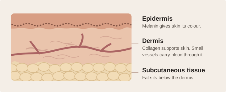
A brown sun spot, a red capillary and a fine line each respond to light in a different way. Choosing a laser starts with understanding which part of the skin we want to change.
What absorbs the light?
Laser light has a chosen wavelength, measured in nanometres (nm). Natural light-absorbing substances in skin are called chromophores. Melanin absorbs light in brown marks. Haemoglobin in blood absorbs light in vessels. Water absorbs the infrared light used for erbium resurfacing.
Why timing matters
Once a target absorbs energy, it can heat up. Choosing a pulse suited to the time the target takes to cool can concentrate the effect there while limiting heat spread. This principle is called selective photothermolysis. Extremely short pulses can also create mechanical stress inside pigment.
How a pico pulse breaks up pigment
“Pico” describes time. One picosecond is one trillionth of a second. A picosecond device emits pulses lasting a fraction of a nanosecond, concentrating energy into a very brief instant.
A pressure wave inside pigment
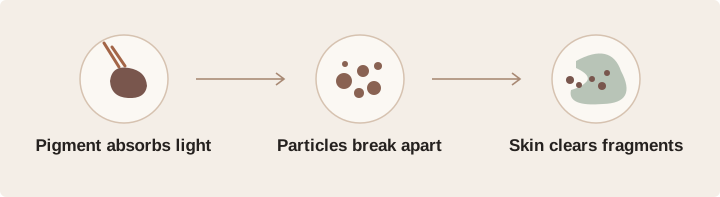
When an absorbing particle takes up energy very quickly, it expands rapidly and creates a pressure wave. This photoacoustic effect can fragment melanin-containing structures or tattoo ink. Heat also contributes. A shorter pulse limits the time available for heat to spread while the pulse is on.
How the colour fades
Superficial pigment can darken briefly, then shed with renewing epidermal cells. Deeper fragments, including some tattoo inks, are taken up by immune cells called macrophages. Clearance is gradual and may be incomplete. A waiting interval lets the skin settle before progress is judged.
Why pico has more than one wavelength
On a suitable Nd:YAG platform, 532 nm green light interacts strongly with superficial melanin. At 1064 nm, near-infrared light is less strongly absorbed by surface melanin and can generally reach deeper targets. The clinician considers pigment colour, depth and the melanin in the surrounding skin.
A different use for fractional pico
A lens array can concentrate energy into many tiny focal points. Above a local threshold, optical breakdown creates microscopic cavities that trigger repair. This is a different delivery pattern from treating a whole sun spot. A small human biopsy study observed different injury patterns with 532 and 1064 nm fractional exposures.
What studies show about pico
These two studies ask different questions. One rates fading in age spots. The other counts how often tattoo ink reached a defined clearance threshold.
Age spots after one treatment
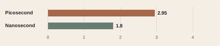
Twenty patients had one side of the face treated with a 532 nm picosecond laser and the other with a 532 nm nanosecond laser. At 12 weeks, three blinded dermatologists graded clearing on a five-point scale.
Tattoo ink reaching a clearance threshold
In a separate randomised trial of 49 patients, investigators treated halves of the same tattoo with different lasers. They counted the halves that reached at least a 75% reduction in colour intensity.
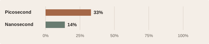
Pico helped some patients in these studies. The results depend on the diagnosis, wavelength, settings and follow-up. These figures cannot predict how a particular age spot, tattoo or case of melasma will respond.
Pico, Nd:YAG and Er:YAG
Pico describes pulse length. Nd:YAG and Er:YAG name the crystals that generate the light. A Nd:YAG laser can itself deliver picosecond pulses, depending on its design.
| Technology | What absorbs the light | How skin can respond |
|---|---|---|
| Picosecond e.g. 532 or 1064 nm | Melanin or ink, depending on its colour, depth and the wavelength. | Very brief pulses can break pigment into smaller particles. Fractional optics can also create small repair zones. |
| Long-pulsed Nd:YAG 1064 nm | Blood in selected dermal vessels. The wavelength can also deliver broader tissue heating. | In a vessel, controlled heating can close its wall. Broader warming can support gradual collagen remodelling. |
| Er:YAG 2940 nm | Water, which is abundant in the skin surface and soft tissues. | Ablative pulses remove thin layers. Gentler pulse trains can build heat while keeping the surface intact. |
How deep does a laser reach?
There is no single depth for a wavelength. Light scatters as it travels. The amount absorbed changes with the target, spot size, water content, skin tone and delivery method. A longer wavelength does not automatically reach farther. At 2940 nm, water absorbs so strongly that most light is deposited close to the surface.
Why a shallow effect can still change the dermis
In fractional Er:YAG resurfacing, narrow columns are treated while adjacent tissue remains intact to support healing. Depth depends in part on energy and passes. Heat and the repair response can influence tissue beyond the surface at which light was first absorbed. This is why treatment depth and the depth of change are different concepts.

Choosing a treatment for your skin
At Skin Cancer Laser Centre in Drummoyne, the most useful starting point is the change you would like to see. A patch of pigment, a visible vessel and a change in texture call for different assessments.
First, identify the concern
A clinician will examine the area and ask when it appeared, how it has changed and what treatments you have tried. A new, changing, bleeding or irregular spot needs medical assessment before a cosmetic laser is considered. Sometimes a biopsy or a different treatment is the next step.
Then, match the treatment to your skin
Your natural skin tone, recent sun exposure, tendency to scar or develop pigmentation, medicines and history of cold sores can change the plan. More melanin in the surrounding skin can also absorb energy. In some circumstances, a test spot and review help assess the response.
Set a realistic recovery window
After pigment treatment, brown spots may darken briefly before fading. Vessel treatment can leave bruising or swelling. With resurfacing, the skin barrier may take longer to heal. Pigment changes, blistering, infection or scarring can occur. Your clinician will explain which risks apply and when to call.
Review progress at the right time
Pigment clearance and collagen repair continue after the initial redness settles. Photographs taken with the same lighting, angle and expression make follow-up more informative. Between visits, follow the care plan and use broad-spectrum SPF 50+ sun protection outdoors.
Discuss your skin with us
Skin Cancer Laser Centre
Unit 12, 50 Victoria Road, Drummoyne NSW 2047
(02) 9719 1600 · info@skincancerlasercentre.com.au